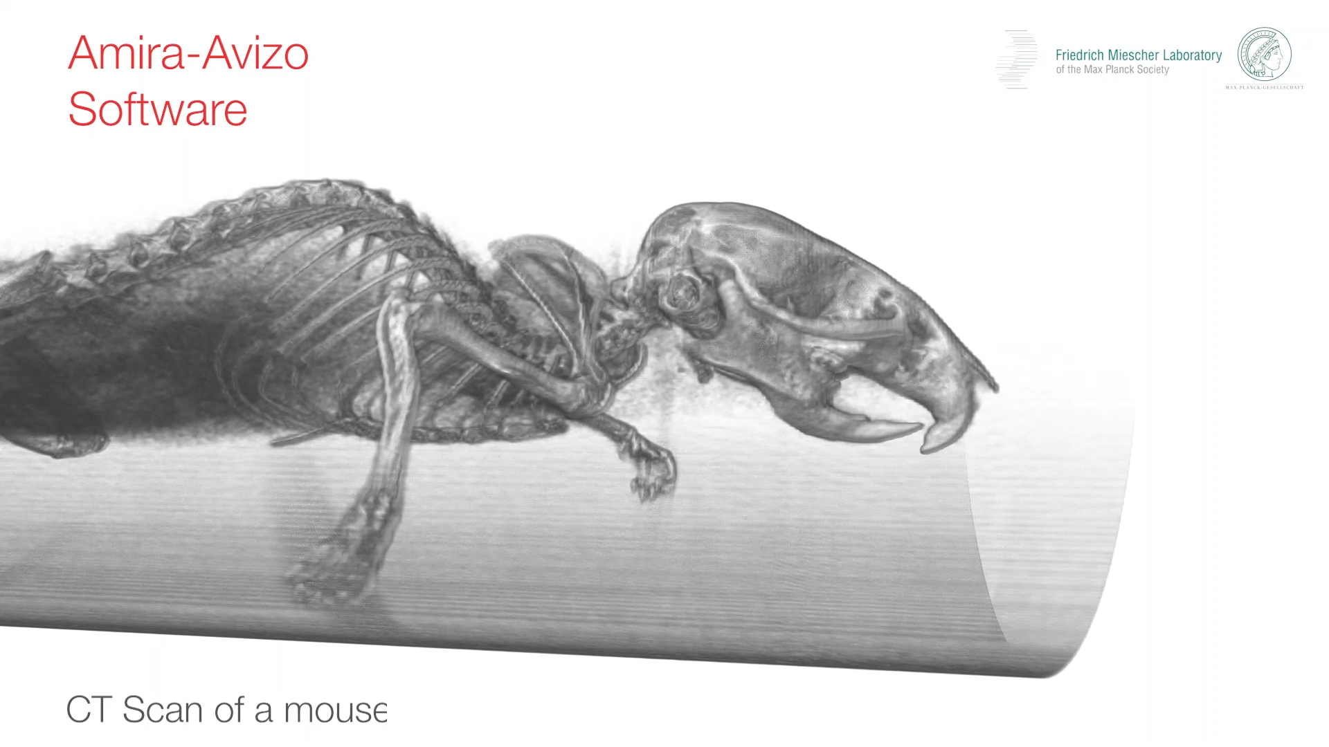
Welcome to the Amira-Avizo Software Use Case Gallery
Below you will find a collection of use cases of our 3D data visualization and analysis software. These use cases include scientific publications, articles, papers, posters, presentations or even videos that show how Amira-Avizo Software is used to address various scientific and industrial research topics.
Use the Domain selector to filter by main application area, and use the Search box to enter keywords related to specific topics you are interested in.
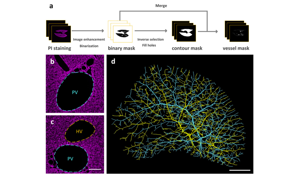
Multiscale reconstruction of various vessels in the intact murine liver lobe
The liver contains a variety of vessels and participates in miscellaneous physiological functions. While past studies generally focused on certain hepatic vessels, we simultaneously obtained all the vessels and cytoarchitectural information of the intact mouse liver lobe at single-cell resolution. […] providing a technology roadmap for studying the fine hepatic vascular structures and their spatial relationship, which will help research into liver diseases and evaluation of medical effi... Read more
Qi Zhang, Anan Li, Siqi Chen, Jing Yuan, Tao Jiang, Xiangning Li, Qingming Luo,Zhao Feng & Hui Gon
High-Resolution Digital Panorama of Multiple Structures in Whole Brain of Alzheimer's Disease Mice
Our study placed emphasis on solving problems in processing high-throughput bright field images and made attempt in developing a method for the extraction and reconstruction of multiple structures. This will facilitate a better understanding of the cerebral anatomical features under the pathological state of AD and shows extensive application prospect in drug efficacy assessment from brain-wide level.
Simultaneously visualizing Amyloid-β (Aβ) plaque with its surrounding brain structu... Read more
Xianzhen Yin, Xiaochuan Zhang, Jingjing Zhang, Weicheng Yang, Xian Sun, Haiyan Zhang, Zhaobing Gao, Hualiang Jiang
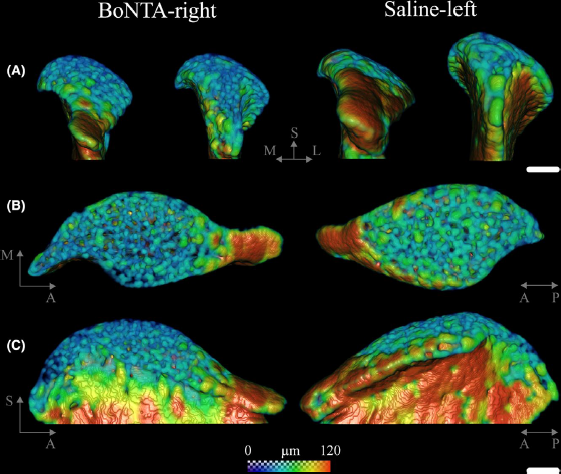
Masseter muscle function influences mandibular bone homeostasis. As previously reported, bone resorption markers increased in the mouse mandibular condyle two days after masseter paralysis induced with botulinum toxin type A (BoNTA), followed by local bone loss.
This study aimed to evaluate the bone quality of both the mandibular condyle and alveolar process in the mandible of adult mice during the early stage of a BoNTA‐induced masseter muscle atrophy, using a combined 3D histomorpho... Read more
Julián Balanta‐Melo, María Angélica Torres‐Quintana, Maximilian Bemmann, Carolina Vega, Constanza González, Kornelius Kupczik, Viviana Toro‐Ibacache, Sonja Buvinic
Loss of adult skeletal muscle stem cells drives age-related neuromuscular junction degeneration
Neuromuscular junction degeneration is a prominent aspect of sarcopenia, the age-associated loss of skeletal muscle integrity. Previously, we showed that muscle stem cells activate and contribute to mouse neuromuscular junction regeneration in response to denervation (Liu et al., 2015). Here, we examined gene expression profiles and neuromuscular junction integrity in aged mouse muscles, and unexpectedly found limited denervation despite a high level of degenerated neuromuscular junctions. In... Read more
Wenxuan Liu, Alanna Klose, Sophie Forman, Nicole D Paris, Lan Wei-LaPierre, Mariela Cortés-Lopéz, Aidi Tan, Morgan Flaherty, Pedro Miura, Robert T Dirksen, Joe V Chakkalakal
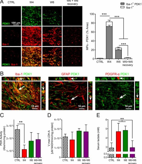
Proinflammatory mononuclear phagocytes (MPs) play a crucial role in the progression of multiple sclerosis (MS) and other neurodegenerative diseases. Despite advances in neuroimaging, there are currently limited available methods enabling noninvasive detection of MPs in vivo. Interestingly, upon activation and subsequent differentiation toward a proinflammatory phenotype MPs undergo metabolic reprogramming that results in increased glycolysis and production of lactate. Hyperpolarized (HP)
Caroline Guglielmetti, Chloé Najac, Alessandro Didonna, Annemie Van der Linden, Sabrina M. Ronen, and Myriam M. Chaumeil
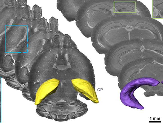
Accumulating evidence indicates the critical importance of cerebrovascular dysfunction in the pathogenesis of Alzheimer’s disease (AD). However, systematic comparative studies on the precise brain vasculature of wild-type and AD model mice are still rare. Using an image optimization method for analyzing Micro-Optical Sectioning Tomography (MOST) data, we generated cross-scale whole-brain 3D atlases that cover the entire vascular system from large vessels down to smallest capillaries at ... Read more
Xiaochuan Zhang, Xianzhen Yin, Jingjing Zhang, Anan Li, Hui Gong, Qingming Luo, Haiyan Zhang, Zhaobing Gao, Hualiang Jiang
Precise Cerebral Vascular Atlas in Stereotaxic Coordinates of Whole Mouse Brain
Understanding amazingly complex brain functions and pathologies requires a complete cerebral vascular atlas in stereotaxic coordinates. Making a precise atlas for cerebral arteries and veins has been a century-old objective in neuroscience and neuropathology. Using micro-optical sectioning tomography (MOST) with a modified Nissl staining method, we acquired five mouse brain data sets containing arteries, veins, and microvessels. Based on the brain-wide vascular spatial structures and brain re... Read more
Benyi Xiong, Anan Li, Yang Lou, Shangbin Chen, Ben Long, Jie Peng, Zhongqin Yang, Tonghui Xu, Xiaoquan Yang, Xiangning Li, Tao Jiang, Qingming Luo and Hui Gong
Multiple membrane extrusion sites drive megakaryocyte migration into bone marrow blood vessels
Platelets, cells central to hemostasis and thrombosis, are formed from parent cell megakaryocytes. Although the process is highly efficient in vivo, our ability to generate them in vitro is still remarkably inefficient. We proposed that greater understanding of the process in vivo is needed and used an imaging approach, intravital correlative light electron microscopy, to visualize platelet generation in bone marrow in the living mouse. In contrast to current understanding, we found that most... Read more
Edward Brown, Leo M Carlin, Claus Nerlov, Cristina Lo Celso, Alastair W Poole
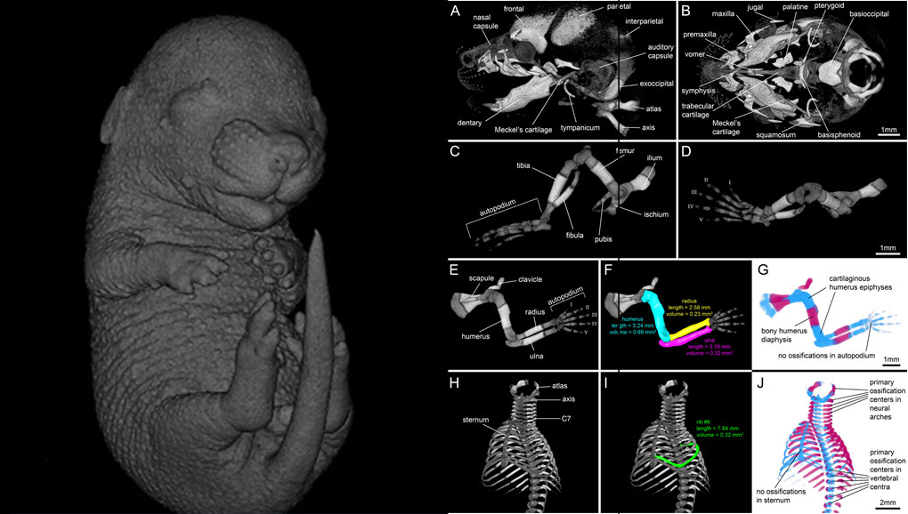
For decades, clearing and staining with Alcian Blue and Alizarin Red has been the gold standard to image vertebrate skeletal development. Here, we present an alternate approach to visualise bone and cartilage based on X-ray microCT imaging, which allows the collection of genuine 3D data of the entire developing skeleton at micron resolution.
Our novel protocol is based on ethanol fixation and staining with Ruthenium Red, and efficiently contrasts cartilage matrix, as demonstrated in wh... Read more
Simone Gabner, Peter Böck, Dieter Fink, Martin Glösmann, Stephan Handschuh
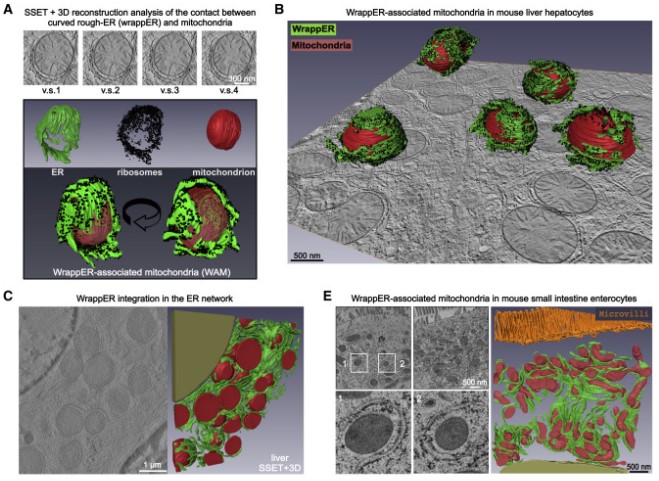
Mitochondria-rough-ER contacts in the liver regulate systemic lipid homeostasis
In this work, we studied mitochondria-rER contacts in vivo by serial section electron tomography (SSET) and 3D reconstruction analysis of cryo-fixed mouse tissue samples. We characterized this inter-organelle association as mitochondria tightly wrapped by sheets of curved rER (wrappER). Further, we used multi-omics and genetic approaches to obtain evidence that the wrappER is a distinct intracellular compartment and demonstrate the importance of wrappER-mitochondria contacts for v... Read more
Irene Anastasia, Nicolò Ilacqua, Andrea Raimondi, Philippe Lemieux, Rana Ghandehari-Alavijeh, Guilhem Faure, Sergei L. Mekhedov, Kevin J. Williams, Federico Caicci, Giorgio Valle, Marta Giacomello, Ariel D. Quiroga, Richard Lehner, Michael J. Miksis, Katalin Toth, Thomas Q. de Aguiar Vallim, Eugene V. Koonin, Luca Scorrano, Luca Pellegrini

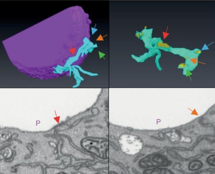
Cell-type specific innervation of cortical pyramidal cells at their apical tufts
We investigated the synaptic innervation of apical tufts of cortical pyramidal cells in a region between layers 1 and 2 using 3-D electron microscopy (3D-EM) applied to four cortical regions in mouse. Across all cortices, we found the relative inhibitory input at the apical dendrite’s main bifurcation to be more than 3-fold stronger for layer 2 pyramidal cells than for all other pyramidal cells. Towards the distal tuft dendrites in upper layer 1, however, the relative inhibitory input was a... Read more
Ali Karimi, Jan Odenthal, Florian Drawitsch, Kevin M. Boergens, Moritz Helmstaedter
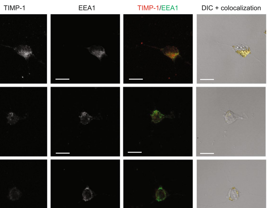
The tissue inhibitor of metalloproteinases-1 (TIMP-1) exerts inhibitory activity against matrix metalloproteinases and cytokine-like effects. We previously showed that TIMP-1 reduces neurite outgrowth in mouse cortical neurons and that this cytokine-like effect depends on TIMP-1 endocytosis mediated by the low-density lipoprotein receptor-related protein-1 (LRP-1). To gain insight into the interaction between TIMP-1 and LRP-1, we considered conformational changes that occur when a ligand bind... Read more
Laurie Verzeaux, Nicolas Belloy, Jessica Thevenard-Devy, Jérôme Devy, Géraldine Ferracci, Laurent Martiny, Stéphane Dedieu, Manuel Dauchez, Hervé Emonard, Nicolas Etique & Emmanuelle Devarenne-Charpentier
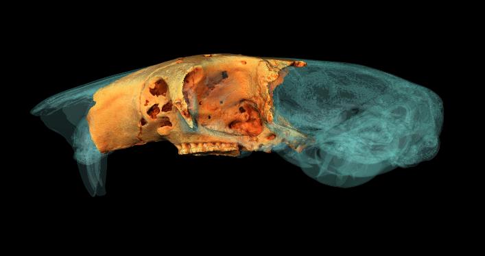
University of York student wins Anatomical Society Best Image Prize using Amira-Avizo Software
PhD student Jesse Hennekam wins for his reconstruction of the skull of a giant dormouse.
A York PhD student has won a prestigious award for his work reconstructing the skull of a giant rodent.
Jesse Hennekam, from the Centre for Anatomical and Human Sciences at the Hull York Medical School, created a digital reconstruction of the skull of a gigantic dormouse (Leithia melitensis), which roamed on the island of Sicily during the Pleistocene, roughly 2 million years ago... Read more
Jesse Hennekam
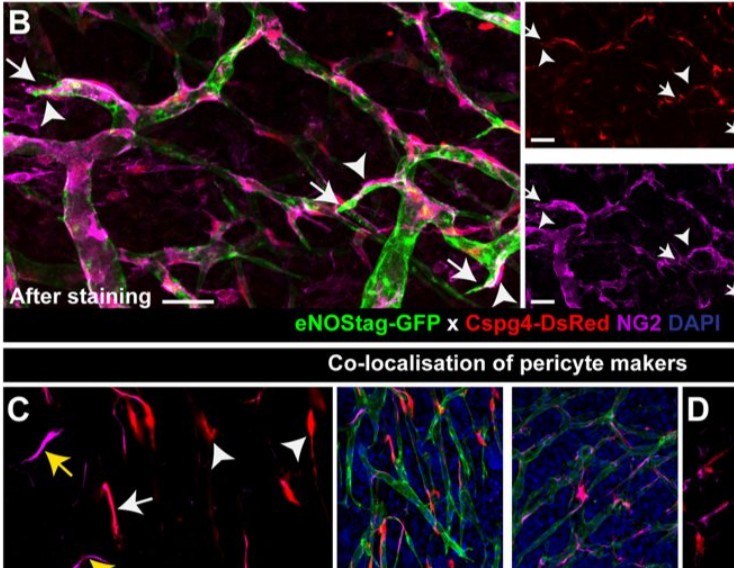
Endothelial cells and pericytes are integral cellular components of the vasculature with distinct interactive functionalities. To study dynamic interactions between these two cells we created two transgenic animal lines. A truncated eNOS (endothelial nitric oxide synthase) construct was used as a GFP tag for endothelial cell evaluation and an inducible Cre-lox recombination, under control of the Pdgfrb (platelet derived growth factor receptor beta) promoter, was created for pericyte assessmen... Read more
Ann L. B. Seynhaeve, Douwe Oostinga, Rien van Haperen, Hanna M. Eilken, Susanne Adams, Ralf H. Adams & Timo L. M. ten Hagen
STIM1 promotes migration, phagosomal maturation and antigen cross-presentation in dendritic cells
Antigen cross-presentation by dendritic cells (DC) stimulates cytotoxic T cell activation to promote immunity to intracellular pathogens, viruses and cancer. Phagocytosed antigens generate potent T cell responses, but the signalling and trafficking pathways regulating their cross-presentation are unclear. Here, we show that ablation of the store-operated-Ca2+-entry regulator STIM1 in mouse myeloid cells impairs cross-presentation and DC migration in vivo and in vitro. Stim1... Read more
Paula Nunes-Hasler, Sophia Maschalidi, Carla Lippens, Cyril Castelbou, Samuel Bouvet, Daniele Guido, Flavien Bermont, Esen Y. Bassoy, Nicolas Page, Doron Merkler, Stéphanie Hugues, Denis Martinvalet, Bénédicte Manoury & Nicolas Demaurex
The Faroe Islands are a group of islands in the North Atlantic that are known for its natural beauty, Viking culture and a special population of house mouse. The Faroese house mouse from the most remote island of the Faroese, Mykines (population: 10 people), looked so distinct when it was discovered that it was declared a subspecies, Mus musculus faeroenesis. These mice are large-bodied and showed an extreme form of left-right asymmetry in its skull. Our research group has... Read more
Yingguang Frank Chan, William H. Beluch, Rémi Blanc
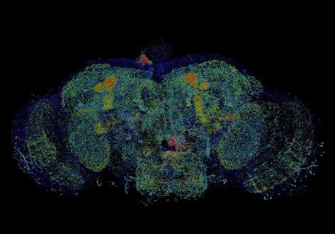
Combined expansion and lattice light sheet microscopy enables high
speed, nanoscale molecular imaging of neural circuits over large volumes.
Optical and electron microscopy have made tremendous inroads in understanding the complexity of the brain, but the former offers insufficient resolution to reveal subcellular details and the latter lacks the throughput and molecular contrast to visualize specific molecular constituents over mm-scale or larger dimensions. We combined expansio... Read more
Ruixuan Gao, Shoh M Asano, Srigokul Upadhyayula, Igor Pisarev, Daniel E Milkie, Tsung-Li Liu, Ved Singh, Austin Graves, Grace H Huynh, Yongxin Zhao, John Bogovic, Jennifer Colonell, Carolyn M Ott, Christopher Zugates, Susan Tappan, Alfredo Rodriguez, Kishore R Mosaliganti, Sean G Megason, Jennifer Lippincott-Schwartz, Adam Hantman, Gerald M Rubin, Tom Kirchhausen, Stephan Saalfeld, Yoshinori Aso, Edward S Boyden, Eric Betzig
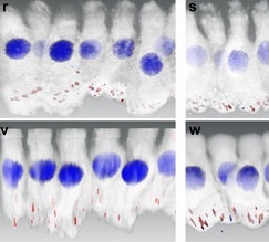
Noise induced hearing loss (NIHL) is a disease that affects millions of Americans. Identifying genetic pathways that influence recovery from noise exposure is an important step forward in understanding NIHL. The transcription factor Foxo3 integrates the cellular response to oxidative stress and plays a role in extending lifespan in many organisms, including humans. Here we show that Foxo3 is required for auditory function after noise exposure in a mouse model system, measured by ABR…Read more
Felicia Gilels, Stephen T. Paquette, Holly J. Beaulac, Anwen Bullen & Patricia M. White
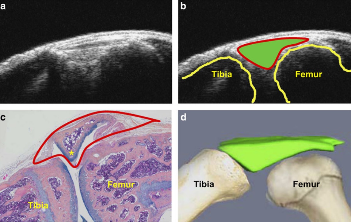
Ultrasound could be a fast and cost-effective means of assessing joint changes in mouse models of posttraumatic osteoarthritis (PTOA). Such models are essential for understanding the biology of this degenerative joint disease and developing new treatments, but noninvasive methods of evaluating disease activity are lacking. Because ultrasound can visualize both joint space volumes and blood flow in the joints, it could provide an alternative to microscopic examination of tissue, assuming it ac... Read more
Hao Xu, Echoe M Bouta, Ronald W Wood, Edward M Schwarz, Yongjun Wang & Lianping Xing